- 簽證留學(xué) |
- 筆譯 |
- 口譯
- 求職 |
- 日/韓語 |
- 德語
The kidneys are located behind the peritoneum in the lumbar region. On the top of each kidney rests an adrenal gland. Each kidney is encased in a capsule of ?brous connective tissue overlaid with fat. An outer-most layer of connective tissue supports the kidney and anchors it to the body wall.
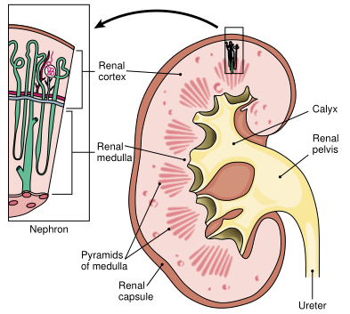
If you look inside the kidney, you will see that it has an outer region, the renal cortex, and an inner region, the renal medulla (Fig. 1). The medulla is divided into triangular sections, each called a pyramid. The pyramids have a lined appearance because they are made up of the loops and collecting tubules of the nephrons, the functional units of the kidney. Each collecting tubule empties into a urine-collecting area called a calyx (from the Latin word meaning "cup"). Several of these smaller minor calyces merge to form a major calyx. The major calyces then unite to form the renal pelvis, the upper funnel-shaped portion of the ureter.

FIGURE 1. Longitudinal section through the kidney showing its internal structure, and an enlarged diagram of a nephron. There are more than 1 million nephrons in each kidney. (Reprinted with permission from Cohen BJ, Wood DL. Memmler's The Human Body in Health and Disease. 9th Ed. Philadelphia: Lippincott Williams & Wilkins, 2000.)
責(zé)任編輯:admin
